Pigmented Mole | Viral Warts | Hyperkeratosis and Skin Lumps

Pigmented Mole

Moles (Nevi) are skin patches caused by an increase in the number of melanocytes, medically referred to as melanocytic nevi. The quantity of moles is influenced by genetic factors and sun exposure. Moles can be smooth or raised, and sometimes hair may grow on their surface, which relates to the depth of melanin accumulation in the skin layers. Moles can appear on various parts of the body, including the face, neck, arms, and back.

Each mole has its own growth pattern; some may enlarge over time, while others may remain unchanged. Moles are generally brown in color, and sometimes may appear flesh-toned. Their color may deepen over the years, or it may fade or even disappear altogether.

Moles are mostly non-cancerous but they could develop into skin cancer (malignant melanoma), SOA doctor consultation is necessary before any treatment.

Types of Nevi (Moles)

nevi are often categorized into flat and raised types based on their appearance. However, in medical terminology, their classification is related to their distribution in different layers of the skin.

The general types of nevi include:

Epidermal Nevi
Pigmentation is located in the epidermal layer, typically appearing smooth with a darker color.

Junctional Nevus
Pigmentation is found between the dermal and epidermal layers, slightly raised, usually brown in color.

Dermal/Intradermal Nevi
Pigmentation is present in the dermal layer, often raised above the skin surface in a dome shape, with a lighter or flesh-colored appearance, and may even have hair.

Preparation Before Mole Removal

The term “mole” generally refers to melanocytic nevi, resulting from the proliferation of melanocytes. Most moles, whether congenital or acquired, are benign. However, melanocytes can undergo malignant transformation, developing into melanoma, a type of skin cancer. Melanoma is often mistaken for a benign mole, and by the time a diagnosis is made, it’s frequently in an advanced, difficult-to-treat stage. Therefore, if a mole exhibits changes in shape, color, size, irregular borders, blurred edges, or unusual texture, immediate medical attention is crucial to rule out malignancy💡. Before attempting mole removal, it’s essential to confirm the mole’s benign nature. If there’s any doubt, a skin biopsy is necessary to ensure it’s not melanoma.

Related Treatments

Mole removal methods vary depending on size, location, and depth.

💡Smaller Moles

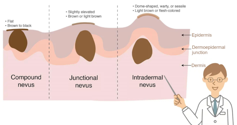
For moles smaller than 0.5cm in diameter, flat or slightly raised, typically located in the epidermis or at the dermis-epidermis junction, laser removal may be considered. This targets superficial tissue, allowing natural healing. Post-treatment redness and pigmentation may occur but fade over time, usually leaving no visible trace or scarring. If the mole’s root is found to be deep during laser treatment, the procedure will be stopped, allowing healing before resuming treatment until completely removed.

💡Larger Moles

Moles larger than 0.5cm or noticeably raised are usually located in the dermis. Because of their deeper roots, laser removal is too traumatic, increasing the risk of large, difficult-to-heal wounds and scarring. Surgical excision is generally preferred, removing the entire mole and its root. This requires stitches, and the scar will fade over time.

Smaller moles leave less noticeable marks. Larger, deeper moles require longer healing times and carry a higher risk of scarring; therefore, early treatment minimizes scarring. The choice between laser removal and surgical excision depends on the physician’s assessment of the mole’s depth and its benign nature. Never attempt mole removal without professional medical advice!

error: Content is protected !!